
AI Endoscopic Depth Assessment for Early Gastric Cancer
10/22/2026
Key Takeaways
- Among resected early gastric cancers in a retrospective cohort at one Japanese hospital, the classifier exceeded its prespecified 75% lower-confidence-bound sensitivity criterion for mucosal disease.
- Specificity for pathologically submucosal disease was lower than overall classification accuracy, and some submucosal cancers received a mucosal label.
- Shallow submucosal lesions were classified correctly less often than deeper lesions, while sensitivity differences by equipment vendor and histology were not statistically significant.
At Okayama University Hospital in Japan, Hirata and colleagues conducted a retrospective, pathology-referenced test of early gastric cancer depth classification in 204 adults with resected EGC: 136 had mucosal cancer and 68 had submucosal cancer. Patients had undergone endoscopic or surgical resection. The test cases followed model development in time but came from the same hospital. Lesion-level classifications were compared with resection histopathology, not treatment outcomes or endoscopists’ unaided assessments.
Deepth EGC uses a Vision Transformer architecture to classify specialist-selected, high-quality white-light still images individually as mucosal or submucosal. Four expert endoscopists selected representative images, with additional investigators verifying the selection; blurred, obscured, or otherwise unsuitable images were excluded. Each lesion contributed at least five images, with a median of eight. A majority vote produced the lesion-level label, and a tie was assigned mucosal. The primary endpoint was mucosal sensitivity, assessed against a prespecified lower bound of a two-sided 95% confidence interval (CI); specificity and accuracy were secondary endpoints.
The classifier identified pathologically mucosal EGC with 91.2% sensitivity (124/136; 95% CI 85.2–94.9%). The lower confidence bound cleared the prespecified criterion. This measure describes recognition of mucosal disease among lesions confirmed as mucosal after resection, not detection of submucosal extension.
Specificity for identifying mucosal cancer was 66.2% (45/68; 95% CI 54.3–76.3%). Here, specificity is the proportion of pathologically submucosal cancers correctly labeled submucosal. It does not establish that the classifier can safely guide treatment selection.
Overall lesion-level classification accuracy was 82.8% (169/204; 95% CI 77.1–87.4%) in a test set randomly sampled from eligible cases to meet a prespecified mix of 136 mucosal and 68 submucosal cancers; accuracy may differ with a different case mix. This measure combines mucosal and submucosal cancers and does not show which direction an incorrect assignment took.
The errors included 12 pathologically mucosal cancers labeled submucosal and 23 pathologically submucosal cancers labeled mucosal. Correct submucosal classification was less frequent for shallow submucosal invasion (SM1) than for deeper submucosal invasion (SM2); those reported subgroups did not encompass every submucosal case. Sensitivity comparisons by endoscope vendor and ulcerative status were not statistically significant. The histology comparison was not statistically significant, but mucosal sensitivity was 78.9% in undifferentiated tumors versus 93.2% in differentiated tumors (P=.07).
The cases were treated in 2018–2021 at the same hospital where the model was developed. The retrospective test used still images chosen for diagnostic suitability rather than unselected images encountered during a live examination, and endoscope vendors were unevenly represented. Neither real-time performance nor AI-assisted versus unaided clinical decisions were evaluated. Errors in either direction could matter when endoscopic resection and surgery are considered, but differences in treatment selection, noncurative resection, surgery, and patient outcomes were not measured. The investigators state that Deepth EGC has medical-device approval in Japan and is commercially available there; they describe it as an aid to, not a replacement for, endoscopist judgment. No U.S. approval can be inferred from the reported Japanese status, which also does not establish clinical benefit.
The investigators report that prospective multicenter validation is underway. Further testing could clarify how the classifier performs with images captured during real-time endoscopy and whether AI-assisted depth assessment changes clinical decisions. For now, its documented performance concerns retrospectively selected white-light still images of resected EGC compared with pathology.
Clinician Questions
How were mucosal and submucosal early gastric cancers confirmed for Deepth EGC evaluation?
Two experienced pathologists independently classified resected early gastric cancer specimens as mucosal or submucosal and resolved disagreements by consensus. Histopathology, rather than Deepth EGC output, supplied the reference diagnosis.
Does Deepth EGC show which image regions influenced its gastric cancer depth prediction?
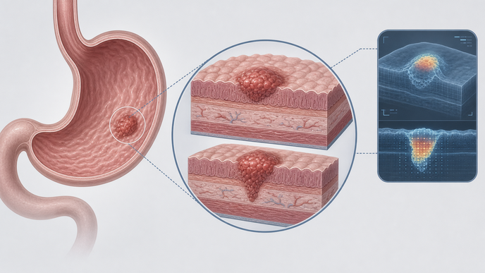
For white-light images of early gastric cancer, Deepth EGC generates probability scores and heat maps highlighting image regions that contributed to its mucosal-versus-submucosal predictions. Those displays were not shown to improve clinical interpretation or treatment choices.